Equine hoof radiograph measurement

Standardize hoof radiograph measurements across your caseload.

DigiHoof helps equine veterinarians measure foot balance and the Lamellar Lucent Zone, review landmarks, compare serial studies, and generate clinical reports.

Veterinarian accounts only Editable landmarks Clinician approval required
Example serial study comparison Left fore · lateral
Reviewed
LLZmid:PCL normalized ratio
9.8% 10.3% +0.5
Palmar angle degrees
3.1° 2.6° −0.5
Sole depth millimetres
14.7 15.5 +0.8

Illustrative values. Every landmark and measurement must be reviewed by the veterinarian.
Serial radiograph measurements

Use the same measurement method at each examination.

Serial comparison requires consistent study identification, calibration, landmark placement, and measurement definitions. DigiHoof records the outputs by horse, limb, view, and examination date.

Defined measurement protocol

Apply named landmarks and calibrated outputs to each suitable radiograph.

Serial comparison

Review structured measurements from the same foot across examination dates.

Clinical documentation

Generate a reviewed measurement report for the medical record and farrier.

Clinical workflow

Process each hoof radiograph study in four steps.

The veterinarian reviews and approves measurements before reporting.

1

Create the study

Record the horse, examination date, limb, and radiographic view.

2

Calibrate and review

Confirm image calibration, then inspect and correct the proposed landmarks.

3

Confirm measurements

Approve the calculated values and retain them as structured study data.

4

Generate the report

Export or share the veterinarian-reviewed measurements and annotations.

Actual clinical workspace

Review the radiograph, landmarks, and report before approval.

These de-identified training studies show the screens used by the veterinarian. Open any image to inspect the full-resolution workspace.

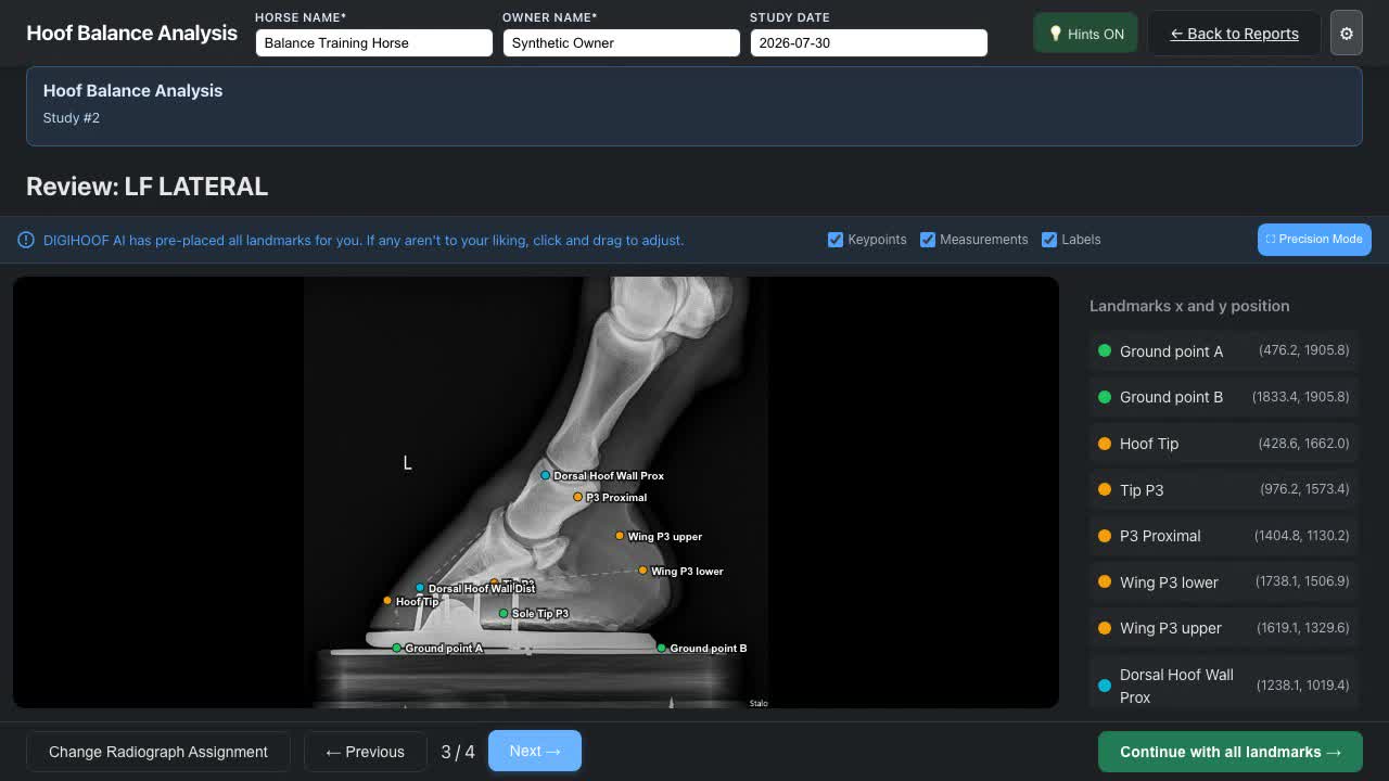
DigiHoof Hoof Balance screen showing a de-identified lateromedial radiograph with editable landmarks
Hoof Balance landmark review Inspect the lateromedial or DP image and correct proposed landmarks before continuing.
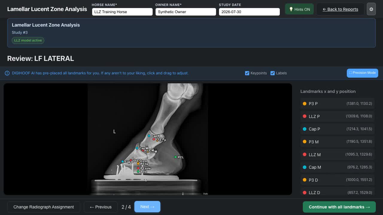
DigiHoof Lamellar Lucent Zone screen showing a de-identified lateromedial radiograph with LLZ landmarks
Lamellar Lucent Zone review Review LLZ, hoof-capsule, P3, sole, and PCL landmarks before accepting the outputs.
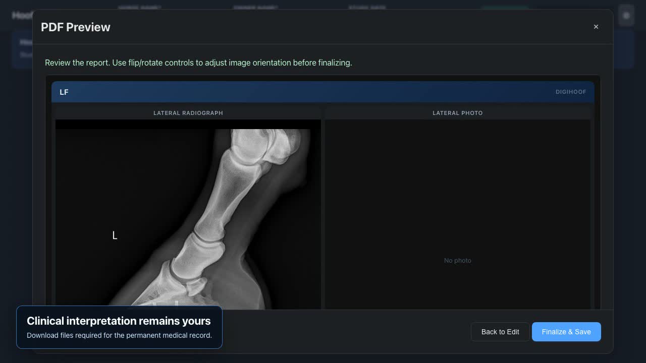
DigiHoof report preview showing a de-identified lateral hoof radiograph before finalization
Report review before finalization Check image orientation and report content before saving the clinical record.

Genuine radiographs; synthetic horse and owner identities. DigiHoof provides measurement assistance and requires veterinarian review.

Three short walkthroughs

See the clinic, Hoof Balance, and LLZ workflows.

The clinical recordings use a de-identified demonstration study with genuine lateral and dorsopalmar radiographs. Captions are included.

Clinic setup · 1 minute

Set up a clinic and invite veterinarians

Create the clinic account, review retention, and send secure individual invitations.

Hoof Balance · 1 minute 9 seconds

Complete a standard Hoof Balance study

Assign radiographs, review editable landmarks, and preview the report.

LLZ · 1 minute 17 seconds

Complete a Lamellar Lucent Zone study

Review LLZ-specific landmarks, dimensions, ratios, and report options.

Available measurements

Hoof Balance and Lamellar Lucent Zone analysis

Both workflows use editable landmarks and retain structured measurement results.

Hoof Balance

Measure calibrated lateral and DP radiographs.

Review foot-balance landmarks and calculated outputs in a consistent workspace, then retain the results for serial comparison.

  • Editable landmarks and outputs
  • Horse, limb, view, and visit context
  • Comparison-ready measurement history
Review Hoof Balance outputs →
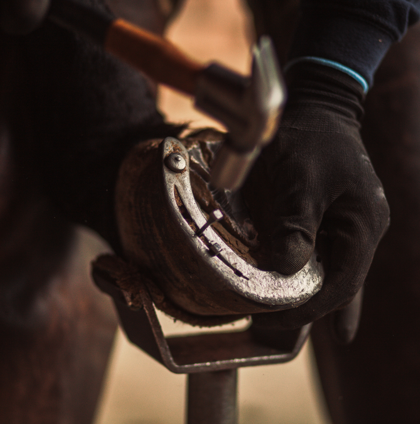
Farrier working on a horse's foot

Clinical reporting

Use reviewed measurements in veterinary–farrier case management.

Reports identify the horse, study date, limb, view, and measurement values. Serial results can support discussion of changes following trimming, shoeing, or medical management.

Review reporting and comparison details →

Data retention

Structured measurements are retained; source files are time-limited.

Source radiographs, photos, reports, and image-bearing share links have clear retention windows. Structured measurements remain available for longitudinal comparison while the clinic account is active.

Review file retention →

For equine veterinarians

Standardize hoof radiograph measurements across your caseload.

Create a veterinarian account to use Hoof Balance. Add the LLZ workflow when required.